Copyright
©2013 Baishideng Publishing Group Co.
World J Gastroenterol. Nov 21, 2013; 19(43): 7726-7734
Published online Nov 21, 2013. doi: 10.3748/wjg.v19.i43.7726
Published online Nov 21, 2013. doi: 10.3748/wjg.v19.i43.7726
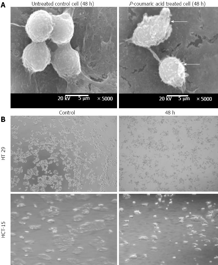
Figure 4 Morphological assessment of p-Coumaric acid treated cells.
A: Human colorectal carcinoma (HCT-15) cells were treated with p-Coumaric acid for 48 h and the cells were observed under scanning electron microscope. Treated cells showed membrane blebbing and shrinkage compared to untreated normal control cells; B: HT 29 and HCT 15 cells were subjected to p-Coumaric acid treatment for 48 h and observed under light microscopy. Treated cells displayed apoptotic features like blebbing and shrinkage compared to untreated normal control cells.
-
Citation: Jaganathan SK, Supriyanto E, Mandal M. Events associated with apoptotic effect of
p -Coumaric acid in HCT-15 colon cancer cells. World J Gastroenterol 2013; 19(43): 7726-7734 - URL: https://www.wjgnet.com/1007-9327/full/v19/i43/7726.htm
- DOI: https://dx.doi.org/10.3748/wjg.v19.i43.7726


