Copyright
©2013 Baishideng Publishing Group Co.
World J Gastroenterol. Oct 28, 2013; 19(40): 6939-6942
Published online Oct 28, 2013. doi: 10.3748/wjg.v19.i40.6939
Published online Oct 28, 2013. doi: 10.3748/wjg.v19.i40.6939
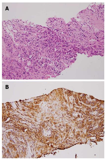
Figure 3 Pathologic findings (hematoxylin/eosin staining) and immunohistochemical staining.
A: The predominant fibrous tissue with necrotic foci and scattered atypical cells characterized by pleomorphic and hyperchromatic nuclei with discohesive arrangement. B: The B-lymphocyte marker (CD20) was strongly positive in the atypical cells.
- Citation: Chen BC, Wang HH, Lin YC, Shih YL, Chang WK, Hsieh TY. Isolated gastric variceal bleeding caused by splenic lymphoma-associated splenic vein occlusion. World J Gastroenterol 2013; 19(40): 6939-6942
- URL: https://www.wjgnet.com/1007-9327/full/v19/i40/6939.htm
- DOI: https://dx.doi.org/10.3748/wjg.v19.i40.6939


