
















©2013 Baishideng Publishing Group Co.
World J Gastroenterol. Oct 28, 2013; 19(40): 6934-6938
Published online Oct 28, 2013. doi: 10.3748/wjg.v19.i40.6934
Published online Oct 28, 2013. doi: 10.3748/wjg.v19.i40.6934
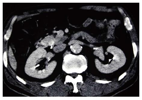
Figure 1 Abdominal computed tomography angiography image of the common bile duct.
Thickening of the wall of the distal common bile duct, which enhances with contrast, was observed (arrowhead).
- Citation: Oshiro Y, Takahashi K, Sasaki R, Kondo T, Sakashita S, Ohkohchi N. Adjuvant surgery for advanced extrahepatic cholangiocarcinoma. World J Gastroenterol 2013; 19(40): 6934-6938
- URL: https://www.wjgnet.com/1007-9327/full/v19/i40/6934.htm
- DOI: https://dx.doi.org/10.3748/wjg.v19.i40.6934