Copyright
©2013 Baishideng Publishing Group Co.
World J Gastroenterol. Jan 28, 2013; 19(4): 597-603
Published online Jan 28, 2013. doi: 10.3748/wjg.v19.i4.597
Published online Jan 28, 2013. doi: 10.3748/wjg.v19.i4.597
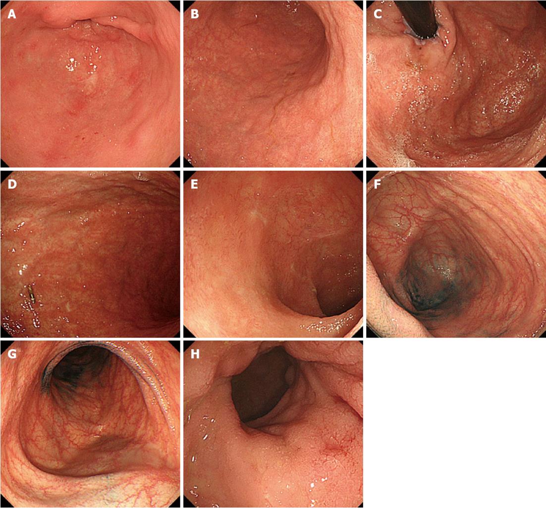
Figure 5 Upper and lower gastrointestinal endoscopic findings 162 d after the steroid and anti-cytomegalovirus therapies.
A: The gastric mucosa in the antrum of the stomach; B: The gastric mucosa in the lower segments of the corpus of the stomach; C: The gastric mucosa in the upper segments of the corpus of the stomach; D: The greater curvature of the stomach near the center. The endoscopic clipping has remained, and nearly the entire mucosa of the stomach has improved to a normal state; E: Normal mucosa in the cecal valve and cecum; F: Normal mucosa in the terminal ileum; G: Marked improvement of the inflammation of the colonic mucosa, and healed ulcers in the sigmoid colon; H: The vascular patterns are now visible, indicating that the normal mucosa has returned in the rectum.
- Citation: Okubo H, Nagata N, Uemura N. Fulminant gastrointestinal graft-versus-host disease concomitant with cytomegalovirus infection: Case report and literature review. World J Gastroenterol 2013; 19(4): 597-603
- URL: https://www.wjgnet.com/1007-9327/full/v19/i4/597.htm
- DOI: https://dx.doi.org/10.3748/wjg.v19.i4.597


