Copyright
©2013 Baishideng Publishing Group Co.
World J Gastroenterol. Jan 28, 2013; 19(4): 536-541
Published online Jan 28, 2013. doi: 10.3748/wjg.v19.i4.536
Published online Jan 28, 2013. doi: 10.3748/wjg.v19.i4.536
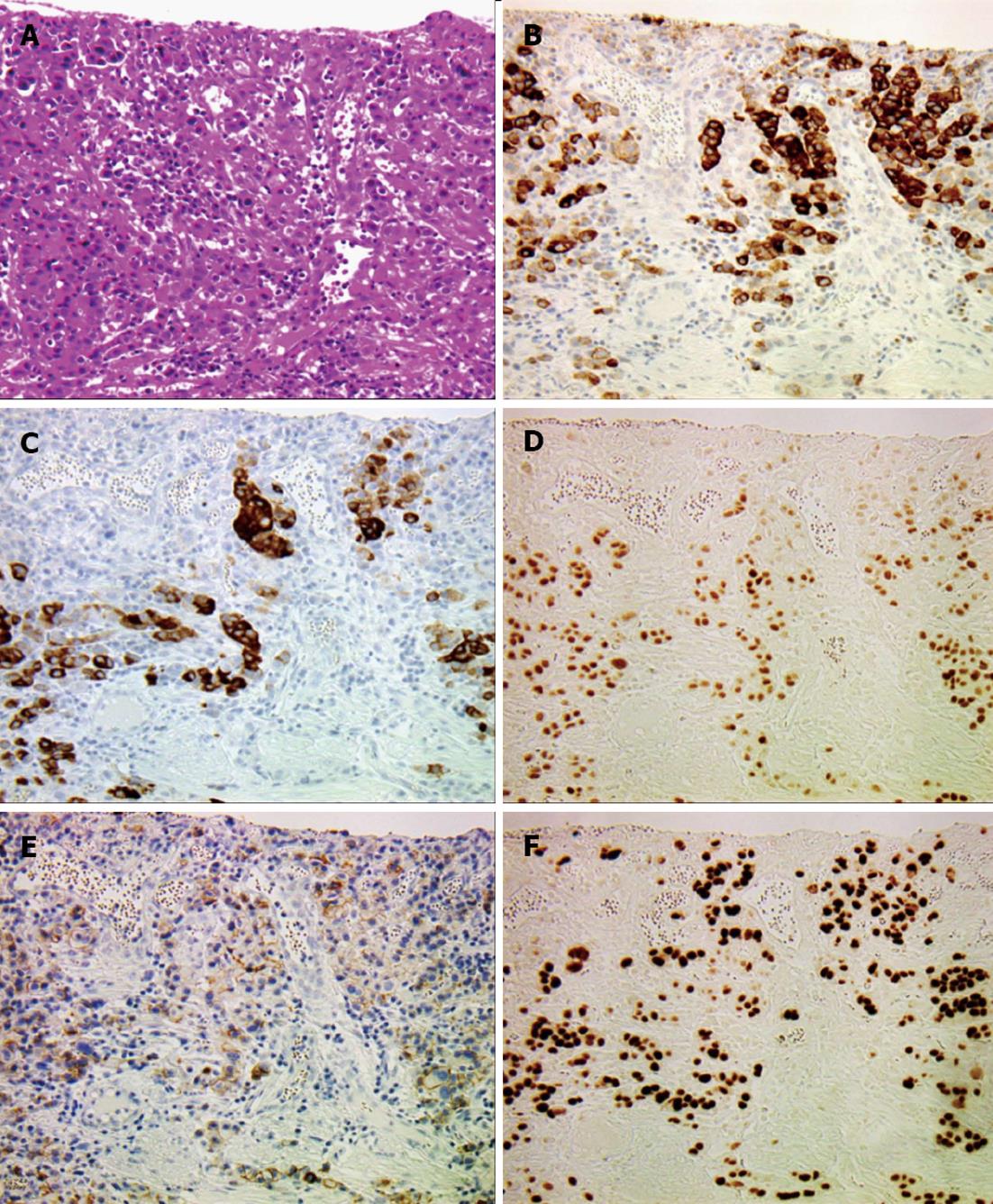
Figure 3 Histological findings of diffuse poorly-differentiated invasive adenocarcinoma (× 200).
A: Hematoxylin and eosin staining of poorly-differentiated invasive adenocarcinoma (PDA). Cancer cells are diffusely scattered with prominent stromal reaction; B: Mucin (MUC) 5AC; C: MUC6; D: Caudal type homebox transcription factor 2 expression is positive in the PDA area; E: E-cad expression is markedly reduced in the PDA area; F: Strong p53 expression is observed in the nuclei of the cells in the PDA area.
- Citation: Makita K, Kitazawa R, Semba S, Fujiishi K, Nakagawa M, Haraguchi R, Kitazawa S. Cdx2 expression and its promoter methylation during metaplasia-dysplasia-carcinoma sequence in Barrett's esophagus. World J Gastroenterol 2013; 19(4): 536-541
- URL: https://www.wjgnet.com/1007-9327/full/v19/i4/536.htm
- DOI: https://dx.doi.org/10.3748/wjg.v19.i4.536


