Copyright
©2013 Baishideng Publishing Group Co.
World J Gastroenterol. Jan 28, 2013; 19(4): 536-541
Published online Jan 28, 2013. doi: 10.3748/wjg.v19.i4.536
Published online Jan 28, 2013. doi: 10.3748/wjg.v19.i4.536
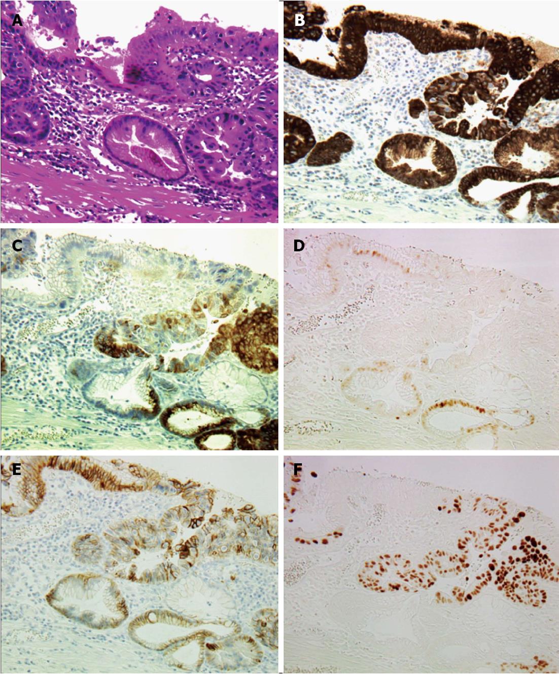
Figure 2 Histological findings of transitional area between intestinal metaplasia and high-grade dysplasia or intramucosal adenocarcinoma (× 200).
A: Hematoxylin and eosin staining of transitional area. Intestinal metaplasia (IM) stretches from the upper left to the lower right corner; B: Mucin (MUC) 5AC immunostaining. Strong MUC5AC expression is observed in both IM and high-grade dysplasia (HD), or intramucosal adenocarcinoma (IMC) areas; C: MUC6 immunostaining. MUC6 expression is observed mostly in parts of the HD or IMC areas; D: Caudal type homebox transcription factor 2 (Cdx2) immunostaining. Cdx2 expression is observed only in the nuclei of the cells in the IM area; E: E-cad immunostaining. E-cad expression is observed on the membranes of cells in both IM and HD or IMC areas; F: p53 immunostaining. Strong p53 expression is observed in the nuclei of the cells in the HD or IMC area.
- Citation: Makita K, Kitazawa R, Semba S, Fujiishi K, Nakagawa M, Haraguchi R, Kitazawa S. Cdx2 expression and its promoter methylation during metaplasia-dysplasia-carcinoma sequence in Barrett's esophagus. World J Gastroenterol 2013; 19(4): 536-541
- URL: https://www.wjgnet.com/1007-9327/full/v19/i4/536.htm
- DOI: https://dx.doi.org/10.3748/wjg.v19.i4.536


