Copyright
©2013 Baishideng Publishing Group Co.
World J Gastroenterol. Oct 14, 2013; 19(38): 6490-6493
Published online Oct 14, 2013. doi: 10.3748/wjg.v19.i38.6490
Published online Oct 14, 2013. doi: 10.3748/wjg.v19.i38.6490
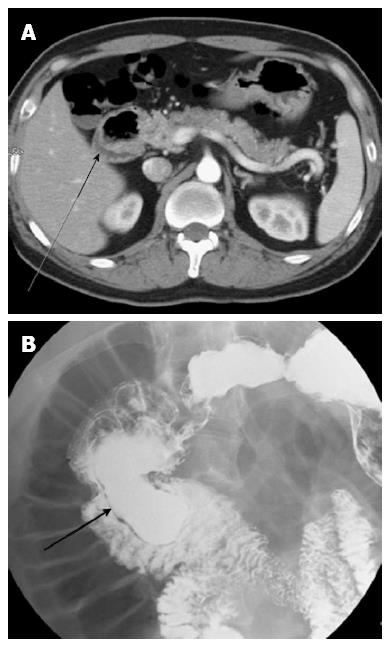
Figure 1 Computed tomography image.
A: A high resolution computed tomography image of the abdomen reveals a circumferential cystic lesion (arrow) in the vicinity of the second part of the duodenum; B: Upper gastrointestinal series showing an large elongated sac-like mass (about 10 cm in length, arrow) arising from the second and proximal third portion of the duodenum.
- Citation: Ko SY, Ko SH, Ha S, Kim MS, Shin HM, Baeg MK. A case of a duodenal duplication cyst presenting as melena. World J Gastroenterol 2013; 19(38): 6490-6493
- URL: https://www.wjgnet.com/1007-9327/full/v19/i38/6490.htm
- DOI: https://dx.doi.org/10.3748/wjg.v19.i38.6490


