Copyright
©2013 Baishideng Publishing Group Co.
World J Gastroenterol. Oct 7, 2013; 19(37): 6310-6314
Published online Oct 7, 2013. doi: 10.3748/wjg.v19.i37.6310
Published online Oct 7, 2013. doi: 10.3748/wjg.v19.i37.6310
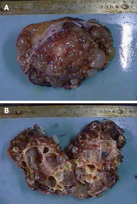
Figure 3 Resected left liver specimen showed a multilocular cystic lesion measuring 15 cm × 9 cm × 8 cm, covered with bullate nodules on the cut surface (A), and opened specimen filled with grayish yellow but clear fluid, the inner surface was smooth without any masses or excrescences (B).
- Citation: Yang ZZ, Li Y, Liu J, Li KF, Yan YH, Xiao WD. Giant biliary cystadenoma complicated with polycystic liver: A case report. World J Gastroenterol 2013; 19(37): 6310-6314
- URL: https://www.wjgnet.com/1007-9327/full/v19/i37/6310.htm
- DOI: https://dx.doi.org/10.3748/wjg.v19.i37.6310


