Copyright
©2013 Baishideng Publishing Group Co.
World J Gastroenterol. Oct 7, 2013; 19(37): 6310-6314
Published online Oct 7, 2013. doi: 10.3748/wjg.v19.i37.6310
Published online Oct 7, 2013. doi: 10.3748/wjg.v19.i37.6310
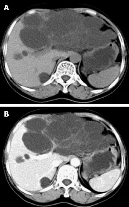
Figure 1 Transverse computed tomography scan showed a left hepatic multiloculated cystic mass measuring 15.
0 cm × 9.1 cm (A) and contrast computed tomography showing enhanced septum of the tumor (B). Simultaneously, multiple sizes of hypoattenuating shadows without enhancement were seen in the right liver lobe.
- Citation: Yang ZZ, Li Y, Liu J, Li KF, Yan YH, Xiao WD. Giant biliary cystadenoma complicated with polycystic liver: A case report. World J Gastroenterol 2013; 19(37): 6310-6314
- URL: https://www.wjgnet.com/1007-9327/full/v19/i37/6310.htm
- DOI: https://dx.doi.org/10.3748/wjg.v19.i37.6310


