Copyright
©2013 Baishideng Publishing Group Co.
World J Gastroenterol. Oct 7, 2013; 19(37): 6299-6303
Published online Oct 7, 2013. doi: 10.3748/wjg.v19.i37.6299
Published online Oct 7, 2013. doi: 10.3748/wjg.v19.i37.6299
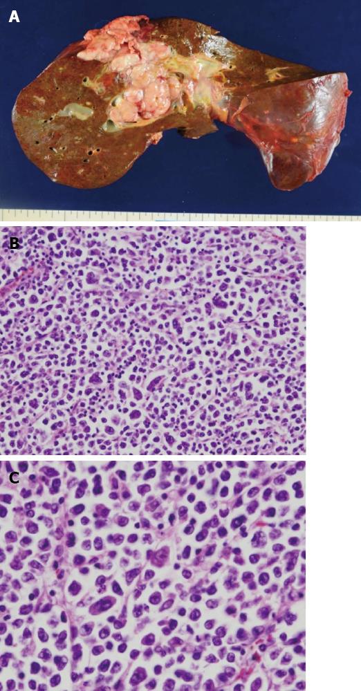
Figure 2 Gross appearance of the primary tumor lesion at autopsy and histological appearance.
A: A large tumor (diameter of 10 cm) is present in the liver. The cut section shows that the tumor is white and solid, with marked necrosis; B: Diffuse infiltration in the liver by monotonous large atypical lymphoid cells (HE staining; original magnification × 200); C: These atypical cells have an abundant basophilic cytoplasm, eccentrically located pleomorphic nuclei, and single, centrally located prominent nucleoli (HE; original magnification × 400).
- Citation: Tani J, Miyoshi H, Nomura T, Yoneyama H, Kobara H, Mori H, Morishita A, Himoto T, Masaki T. A case of plasmablastic lymphoma of the liver without human immunodeficiency virus infection. World J Gastroenterol 2013; 19(37): 6299-6303
- URL: https://www.wjgnet.com/1007-9327/full/v19/i37/6299.htm
- DOI: https://dx.doi.org/10.3748/wjg.v19.i37.6299


