Copyright
©2013 Baishideng Publishing Group Co.
World J Gastroenterol. Sep 28, 2013; 19(36): 6110-6113
Published online Sep 28, 2013. doi: 10.3748/wjg.v19.i36.6110
Published online Sep 28, 2013. doi: 10.3748/wjg.v19.i36.6110
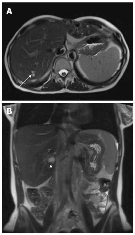
Figure 6 Follow up magnetic resonance imaging axial (A) and coronal (B) balanced gradient echo sequences (True FISP; Siemens Medical, Erlangen, Germany) through the liver performed as part of routine annual tumor surveillance at 1 year following venoplasty of the hepatic vein and inferior vena cava stenosis demonstrated normal appearance of the liver apart from simple parenchymal cysts (arrow).
No ascites was seen.
- Citation: Strovski E, Liu D, Scudamore C, Ho S, Yoshida E, Klass D. Magnetic resonance venography and liver transplant complications. World J Gastroenterol 2013; 19(36): 6110-6113
- URL: https://www.wjgnet.com/1007-9327/full/v19/i36/6110.htm
- DOI: https://dx.doi.org/10.3748/wjg.v19.i36.6110


