Copyright
©2013 Baishideng Publishing Group Co.
World J Gastroenterol. Sep 28, 2013; 19(36): 6011-6019
Published online Sep 28, 2013. doi: 10.3748/wjg.v19.i36.6011
Published online Sep 28, 2013. doi: 10.3748/wjg.v19.i36.6011
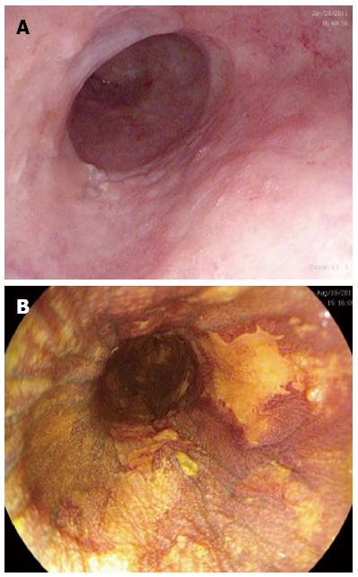
Figure 1 Endoscopic views of a segment of squamous high grade dysplasia with macroscopic lesions suggestive of dysplasia with (A) high definition white light endoscopy and confirmation of areas of dysplasia with (B) Lugol’s iodine dye spray representing unstained lesions.
- Citation: Haidry RJ, Butt MA, Dunn J, Banks M, Gupta A, Smart H, Bhandari P, Smith LA, Willert R, Fullarton G, John M, Pietro MD, Penman I, Novelli M, Lovat LB. Radiofrequency ablation for early oesophageal squamous neoplasia: Outcomes form United Kingdom registry. World J Gastroenterol 2013; 19(36): 6011-6019
- URL: https://www.wjgnet.com/1007-9327/full/v19/i36/6011.htm
- DOI: https://dx.doi.org/10.3748/wjg.v19.i36.6011


