Copyright
©2013 Baishideng Publishing Group Co.
World J Gastroenterol. Sep 7, 2013; 19(33): 5473-5484
Published online Sep 7, 2013. doi: 10.3748/wjg.v19.i33.5473
Published online Sep 7, 2013. doi: 10.3748/wjg.v19.i33.5473
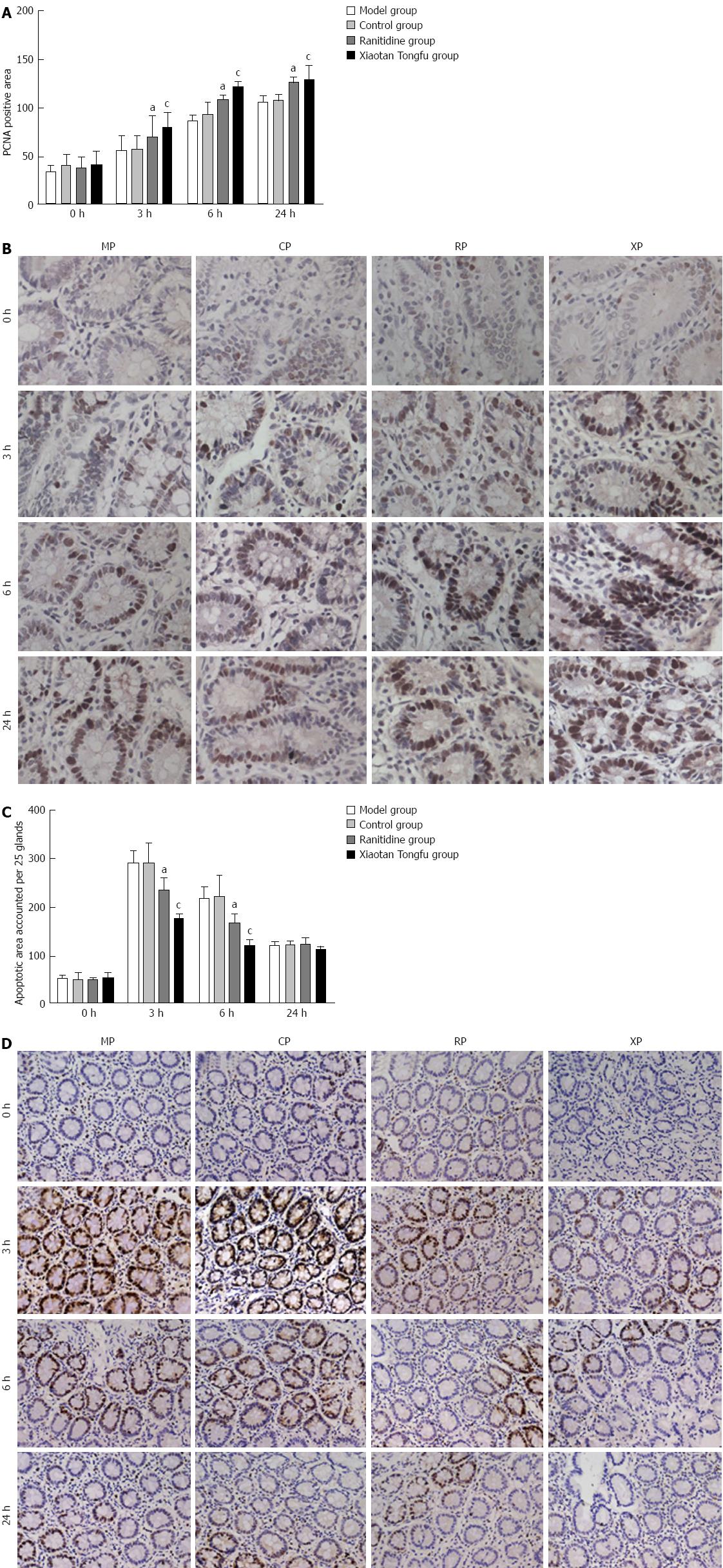
Figure 6 Measurement of cell proliferation and mucosal cell apoptosis (n = 6 for each group).
A: The cell proliferation was significantly different between the ranitidine (RP) and Xiaotan Tongfu granule (XP) groups (P < 0.05) at 3 and 6 h; B: Proliferating cell nuclear antigen (PCNA) immunoreactivity was observed in the gastric surface epithelium, and this staining was focused in the nucleus; C, D: Strongly apoptotic cells were observed in the nucleus of the gastric surface epithelium. Similar to the cell proliferation, the cell apoptosis was significantly different between the RP and XP groups at 3 and 6 h (P < 0.05). Original magnification × 400. aP < 0.05 vs the model group (MP group); cP < 0.05 vs the control group (CP group).
- Citation: Yan B, Shi J, Xiu LJ, Liu X, Zhou YQ, Feng SH, Lv C, Yuan XX, Zhang YC, Li YJ, Wei PK, Qin ZF. Xiaotan Tongfu granules contribute to the prevention of stress ulcers. World J Gastroenterol 2013; 19(33): 5473-5484
- URL: https://www.wjgnet.com/1007-9327/full/v19/i33/5473.htm
- DOI: https://dx.doi.org/10.3748/wjg.v19.i33.5473


