Copyright
©2013 Baishideng Publishing Group Co.
World J Gastroenterol. Aug 21, 2013; 19(31): 5187-5194
Published online Aug 21, 2013. doi: 10.3748/wjg.v19.i31.5187
Published online Aug 21, 2013. doi: 10.3748/wjg.v19.i31.5187
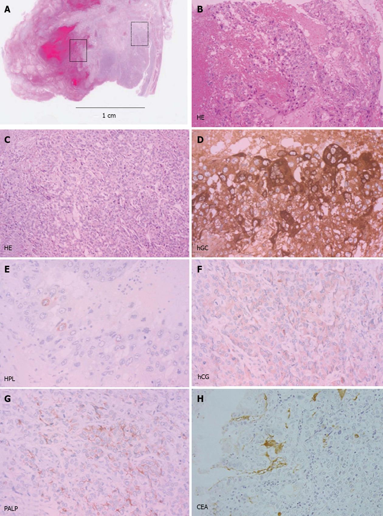
Figure 2 Pathological findings (hematoxylin/eosin staining and immunohistochemical staining).
A: The tumor had two components, as indicated by small boxes with solid and dashed lines. Hematoxylin/eosin (HE) × 40; B: In the area marked by a solid line in panel A, unusual multinucleated giant cells in a characteristic dimorphic plexiform pattern associated with hemorrhage and necrosis were observed. HE × 100; C: Atypical mononucleated cells demonstrated a solid and sheet growth pattern in the area marked by a dashed line in panel A. HE × 100; D, E: Tumor cells were diffusely positive for β-human chorionic gonadotropin (hCG) and focally positive for human placental lactogen (HPL) in the area marked by a solid line in panel A. HE × 200; F, G: The tumor cells were positive for beta-human chorionic gonadotropin and placental alkaline phosphatase (PALP) in the area marked by a dashed line in panel A. HE × 200; H: Immunoreactivity was focally positive for carcinoembryonic antigen (CEA). HE × 100.
- Citation: Takahashi K, Tsukamoto S, Saito K, Ohkohchi N, Hirayama K. Complete response to multidisciplinary therapy in a patient with primary gastric choriocarcinoma. World J Gastroenterol 2013; 19(31): 5187-5194
- URL: https://www.wjgnet.com/1007-9327/full/v19/i31/5187.htm
- DOI: https://dx.doi.org/10.3748/wjg.v19.i31.5187


