Copyright
©2013 Baishideng Publishing Group Co.
World J Gastroenterol. Aug 21, 2013; 19(31): 5174-5177
Published online Aug 21, 2013. doi: 10.3748/wjg.v19.i31.5174
Published online Aug 21, 2013. doi: 10.3748/wjg.v19.i31.5174
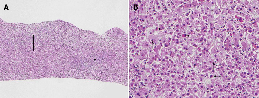
Figure 1 Pathological liver tissue.
A: Diffuse portal and lobular inflammatory cell infiltrates (long arrows) [hematoxylin and eosin (HE), × 20]; B: Hepatocytes are reactive with prominent ballooning degeneration (short arrows) and individual cell necrosis (long arrows) (HE, × 200).
- Citation: Patel SS, Beer S, Kearney DL, Phillips G, Carter BA. Green tea extract: A potential cause of acute liver failure. World J Gastroenterol 2013; 19(31): 5174-5177
- URL: https://www.wjgnet.com/1007-9327/full/v19/i31/5174.htm
- DOI: https://dx.doi.org/10.3748/wjg.v19.i31.5174


