Copyright
©2013 Baishideng Publishing Group Co.
World J Gastroenterol. Aug 14, 2013; 19(30): 5006-5010
Published online Aug 14, 2013. doi: 10.3748/wjg.v19.i30.5006
Published online Aug 14, 2013. doi: 10.3748/wjg.v19.i30.5006
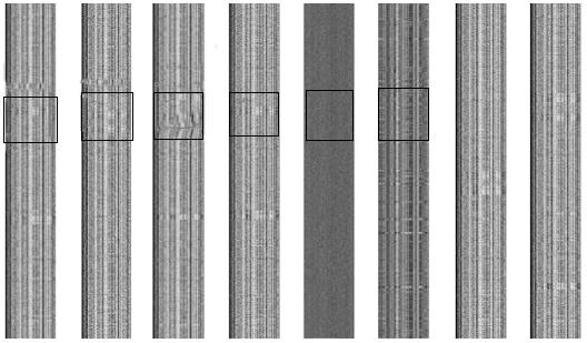
Figure 1 2-D barcode images of genomes of Helicobacter pylori strains J99, G27, 26695, HPAG1, P12, and Shi470, Escherichia coli O157:H7 strain EDL933, and a random sequence.
The y-axis represents the genome axis from top-down, with each pixel representing a fragment of n = 1000 bp; the x-axis represents the 4-nucletide frequencies. The abnormal barcode regions are demarcated by a rectangle.
-
Citation: Wang GQ, Xu JT, Xu GY, Zhang Y, Li F, Suo J. Predicting a novel pathogenicity island in
Helicobacter pylori by genomic barcoding. World J Gastroenterol 2013; 19(30): 5006-5010 - URL: https://www.wjgnet.com/1007-9327/full/v19/i30/5006.htm
- DOI: https://dx.doi.org/10.3748/wjg.v19.i30.5006


