
















©2013 Baishideng Publishing Group Co.
World J Gastroenterol. Jan 21, 2013; 19(3): 422-425
Published online Jan 21, 2013. doi: 10.3748/wjg.v19.i3.422
Published online Jan 21, 2013. doi: 10.3748/wjg.v19.i3.422
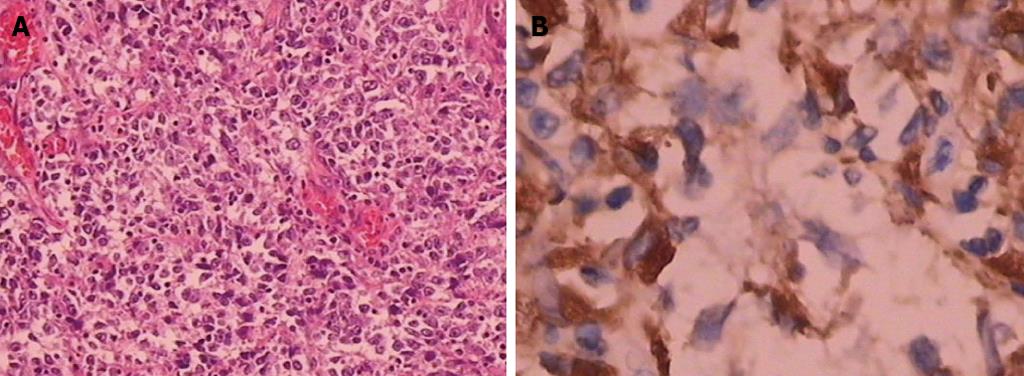
Figure 5 Histopathologic exam showed the tumor cells were marked inequality in the sizes, the cytoplasm was abundant and eosinophilic, the nuclei were large, round to oval, bizarre multinuclear giant cells were scattered.
The tumor cells expressed CD68. A: Hematoxylin and eosin ×100; B: Immunohistochemical staining for CD68 ×400.
- Citation: Shen XZ, Liu F, Ni RJ, Wang BY. Primary histiocytic sarcoma of the stomach: A case report with imaging findings. World J Gastroenterol 2013; 19(3): 422-425
- URL: https://www.wjgnet.com/1007-9327/full/v19/i3/422.htm
- DOI: https://dx.doi.org/10.3748/wjg.v19.i3.422