Copyright
©2013 Baishideng Publishing Group Co.
World J Gastroenterol. Jan 21, 2013; 19(3): 418-421
Published online Jan 21, 2013. doi: 10.3748/wjg.v19.i3.418
Published online Jan 21, 2013. doi: 10.3748/wjg.v19.i3.418
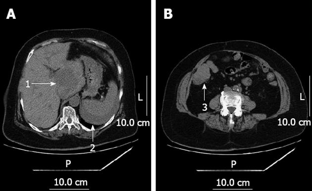
Figure 1 Urgent computed tomography.
A: Sagittal view; B: Transverse view. 1: A ruptured mass of 6 cm × 6 cm × 6 cm localized in the I hepatic segment; 2: Perisplenic hemoperitoneum; 3: A 5 cm × 5 cm × 5 cm mass abutting the ileocecus without lymph node enlargement. L: Left; P: Posterior.
- Citation: Sun LH, Han HQ, Wang PZ, Tian WJ. Emergency caudate lobectomy for ruptured hepatocellular carcinoma with multiple primary cancers. World J Gastroenterol 2013; 19(3): 418-421
- URL: https://www.wjgnet.com/1007-9327/full/v19/i3/418.htm
- DOI: https://dx.doi.org/10.3748/wjg.v19.i3.418


