Copyright
©2013 Baishideng Publishing Group Co.
World J Gastroenterol. Aug 7, 2013; 19(29): 4836-4840
Published online Aug 7, 2013. doi: 10.3748/wjg.v19.i29.4836
Published online Aug 7, 2013. doi: 10.3748/wjg.v19.i29.4836
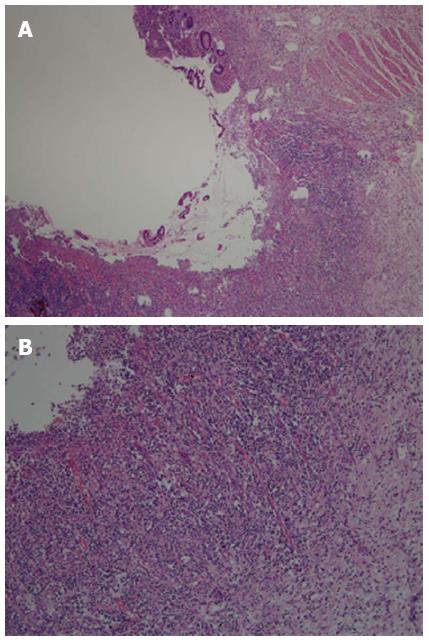
Figure 5 Histological examination.
A: Glands of the small intestinal mucosa were necrotic and exfoliated, with intestinal villous atrophy and ulcer formation [hematoxylin and eosin (HE) stain, × 40]; B: Extensive small lymphocytes and plasma cell infiltration in the lamina propria, submucosa, and serosa of the terminal ileum (HE stain, × 100).
- Citation: Gao X, Wang ZJ. Idiopathic chronic ulcerative enteritis with perforation and recurrent bleeding: A case report. World J Gastroenterol 2013; 19(29): 4836-4840
- URL: https://www.wjgnet.com/1007-9327/full/v19/i29/4836.htm
- DOI: https://dx.doi.org/10.3748/wjg.v19.i29.4836


