Copyright
©2013 Baishideng Publishing Group Co.
World J Gastroenterol. Jul 21, 2013; 19(27): 4437-4442
Published online Jul 21, 2013. doi: 10.3748/wjg.v19.i27.4437
Published online Jul 21, 2013. doi: 10.3748/wjg.v19.i27.4437
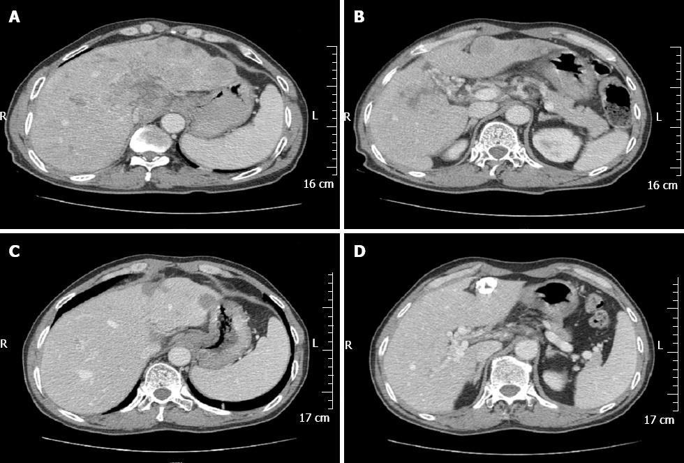
Figure 2 Computed tomography imaging of the gastric tumor and liver metastatic tumor.
A and B: Computed tomography-scan revealing thickening of the wall of antrum, enlarged lymph nodes at the lesser curvature, multiple hepatic tumors in the bilateral lobes of the liver, tumor thrombus in the portal vein and its branches; C and D: After comprehensive therapies, enlarged lymph nodes had decreased in size and the liver metastatic foci were stable.
- Citation: Ye MF, Tao F, Liu F, Sun AJ. Hepatoid adenocarcinoma of the stomach: A report of three cases. World J Gastroenterol 2013; 19(27): 4437-4442
- URL: https://www.wjgnet.com/1007-9327/full/v19/i27/4437.htm
- DOI: https://dx.doi.org/10.3748/wjg.v19.i27.4437


