Copyright
©2013 Baishideng Publishing Group Co.
World J Gastroenterol. Jul 14, 2013; 19(26): 4106-4118
Published online Jul 14, 2013. doi: 10.3748/wjg.v19.i26.4106
Published online Jul 14, 2013. doi: 10.3748/wjg.v19.i26.4106
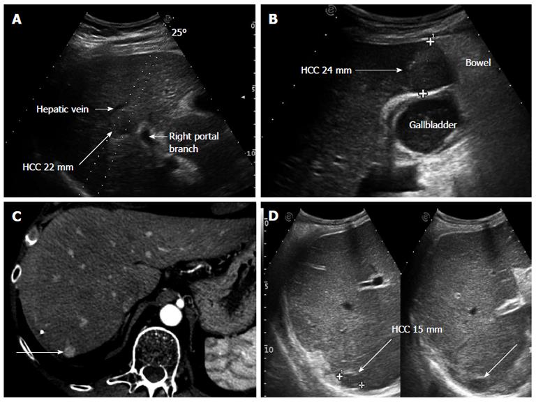
Figure 1 Clinical cases in which performing hepatic resection or radiofrequency ablation had to be decided.
A: Small hepatocellular carcinoma (HCC), 22 mm in diameter, located centrally in the right liver lobe in a patient with MELD 10 and clinical signs of portal hypertension. Surgery would have required a right hepatectomy, thus, radiofrequency ablation was preferred even if a reduced rate of complete necrosis could be expected due to the possible heat sink effect of the nearby large vessels; B: The tumor is located sub-capsular, close to the bowel loops and in strict contact with the gallbladder, implying various technical contraindications to percutaneous ablation. Open surgery was the strategy adopted; C: The tumor (long arrow), shown in the arterial phase of contrast enhancement at computed tomography scan, is located sub-capsular at the liver dome; D: Ultrasonography confirms the tumor (long arrow) to lie very deep and without a safe needle track; in fact, these images are taken in deep inspiration, the lesion being hardly visible during normal breathing. The location was considered to contraindicate percutaneous ablation and surgery was performed.
-
Citation: Cucchetti A, Piscaglia F, Cescon M, Ercolani G, Pinna AD. Systematic review of surgical resection
vs radiofrequency ablation for hepatocellular carcinoma. World J Gastroenterol 2013; 19(26): 4106-4118 - URL: https://www.wjgnet.com/1007-9327/full/v19/i26/4106.htm
- DOI: https://dx.doi.org/10.3748/wjg.v19.i26.4106


