Copyright
©2013 Baishideng Publishing Group Co.
World J Gastroenterol. Jun 28, 2013; 19(24): 3831-3840
Published online Jun 28, 2013. doi: 10.3748/wjg.v19.i24.3831
Published online Jun 28, 2013. doi: 10.3748/wjg.v19.i24.3831
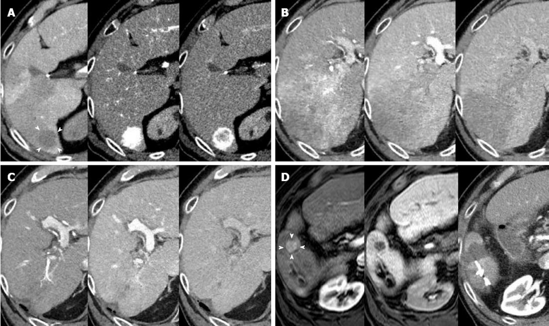
Figure 4 Representative follow-up images of successfully treated hepatocellular carcinoma in an elderly patient.
A: A classical hepatocellular carcinoma (HCC) was detected in segment 6 of the liver on April 12, 2009 as demonstrated by (1) a lower intensity up on computed tomography (CT) during arterial portography, indicated by arrowheads (left); (2) vigorous staining during the arterial phase of the CT during hepatic arteriography (middle); and (3) washout with a corona-like peripheral enhancement during the equilibrium phase of the CT during hepatic arteriography (right); B: A dynamic CT 25 mo after the initial radiofrequency ablation revealing recurrent HCCs, which had spread to large areas of segments 6 and 7 and extended to the main trunk of the right portal vein. The images were obtained during arterial, portal and equilibrium phases of the dynamic CT study, shown in order from left to right; C: After hepatic arterial infusion chemotherapy via a catheter using 5-fluorouracil and cis-diamminedichloroplatinum for 15 mo, an enormous tumor reduction, including the portal vein tumor thrombus, was achieved. The images were obtained during arterial, portal, and equilibrium phases of the dynamic CT study, shown in order from left to right; D: A new 10-mm lesion appeared in segment 6 during hepatic arterial infusion chemotherapy treatment, and a second radiofrequency ablation (RFA) was applied 41 mo after the initial RFA. Magnetic resonance imaging study using a contrast medium of gadolinium ethoxybenzyl diethylene-triamine-pentaacetic-acid showing the arterial supply (left, arrowheads) and a defect in the hepatobiliary phase (middle) of the study. An arterial phase image of the dynamic CT (right) obtained one day after RFA revealing that the ablated area of lower intensity included the target.
- Citation: Suda T, Nagashima A, Takahashi S, Kanefuji T, Kamimura K, Tamura Y, Takamura M, Igarashi M, Kawai H, Yamagiwa S, Nomoto M, Aoyagi Y. Active treatments are a rational approach for hepatocellular carcinoma in elderly patients. World J Gastroenterol 2013; 19(24): 3831-3840
- URL: https://www.wjgnet.com/1007-9327/full/v19/i24/3831.htm
- DOI: https://dx.doi.org/10.3748/wjg.v19.i24.3831


