Copyright
©2013 Baishideng Publishing Group Co.
World J Gastroenterol. Jun 28, 2013; 19(24): 3747-3760
Published online Jun 28, 2013. doi: 10.3748/wjg.v19.i24.3747
Published online Jun 28, 2013. doi: 10.3748/wjg.v19.i24.3747
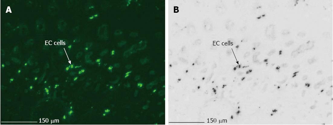
Figure 3 Photomicrographs of immunohistologically stained 8 μm sections of pig gastric fundus showing.
A: MAB352 (1:200) immunofluorescent enterochromaffin cells (EC) in the epithelium (× 20); B: Invert color visualization of MAB352 (1:200) immunofluorescent EC cells.
- Citation: Priem EK, Maeyer JHD, Vandewoestyne M, Deforce D, Lefebvre RA. Predominant mucosal expression of 5-HT4(+h) receptor splice variants in pig stomach and colon. World J Gastroenterol 2013; 19(24): 3747-3760
- URL: https://www.wjgnet.com/1007-9327/full/v19/i24/3747.htm
- DOI: https://dx.doi.org/10.3748/wjg.v19.i24.3747


