Copyright
©2013 Baishideng Publishing Group Co.
World J Gastroenterol. Jun 21, 2013; 19(23): 3583-3595
Published online Jun 21, 2013. doi: 10.3748/wjg.v19.i23.3583
Published online Jun 21, 2013. doi: 10.3748/wjg.v19.i23.3583
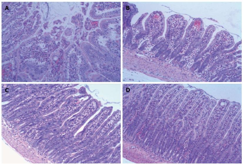
Figure 4 Histopathology of ileum sections of different groups at 6 h after ischemia/reperfusion injury (hematoxylin and eosin, × 100).
A: In the ischemia/reperfusion (I/R) injury group there was marked intestinal mucosa injury at 6 h, with intestinal mucosa degradation and disintegration of the lamina propria, hemorrhage, and ulceration. B: In the bone-marrow mesenchymal stem cells + I/R injury group at 6 h, the damaged mucosa had recovered and there was extension of the subepithelial space with moderate lifting of the epithelial layer from the lamina propria, massive epithelial lifting down the sides of the villi, and ulceration at the villous tips. C and D: In the anti-tumor necrosis factor (TNF)-α + I/R injury group and the anti-TNF-αR1-IgG + I/R injury group at 6 h, the damaged mucosa had almost recovered to resemble that in the Sham control group.
-
Citation: Shen ZY, Zhang J, Song HL, Zheng WP. Bone-marrow mesenchymal stem cells reduce rat intestinal ischemia-reperfusion injury, ZO-1 downregulation and tight junction disruption
via a TNF-α-regulated mechanism. World J Gastroenterol 2013; 19(23): 3583-3595 - URL: https://www.wjgnet.com/1007-9327/full/v19/i23/3583.htm
- DOI: https://dx.doi.org/10.3748/wjg.v19.i23.3583


