Copyright
©2013 Baishideng Publishing Group Co.
World J Gastroenterol. Jun 21, 2013; 19(23): 3583-3595
Published online Jun 21, 2013. doi: 10.3748/wjg.v19.i23.3583
Published online Jun 21, 2013. doi: 10.3748/wjg.v19.i23.3583
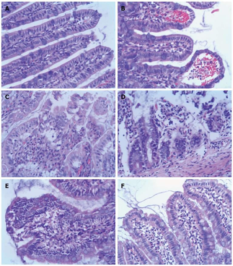
Figure 3 Histopathology of ileum sections at different time points after intestinal ischemia/reperfusion injury (hematoxylin and eosin, × 200).
A: Sham group; the intestine showed normal villous architecture and glands, with no vascular congestion; B: In the ischemia/reperfusion (I/R) injury 2 h group, the degree of intestinal mucosa injury was marked with massive epithelial lifting down the sides of the villi and ulceration at the villous tips; C: At 6 h, there was intestinal mucosa degradation and disintegration of the lamina propria, hemorrhage, and ulceration; D: At 24 h, the damaged mucosa showed denuded villi with dilated capillaries and increased cellularity of the lamina propria; E: In the I/R injury group at 72 h, there was massive epithelial lifting down the sides of the villi and ulceration at the villous tips; F: However, at 144 h, the damaged mucosa had recovered.
-
Citation: Shen ZY, Zhang J, Song HL, Zheng WP. Bone-marrow mesenchymal stem cells reduce rat intestinal ischemia-reperfusion injury, ZO-1 downregulation and tight junction disruption
via a TNF-α-regulated mechanism. World J Gastroenterol 2013; 19(23): 3583-3595 - URL: https://www.wjgnet.com/1007-9327/full/v19/i23/3583.htm
- DOI: https://dx.doi.org/10.3748/wjg.v19.i23.3583


