Copyright
©2013 Baishideng Publishing Group Co.
World J Gastroenterol. Jun 14, 2013; 19(22): 3517-3519
Published online Jun 14, 2013. doi: 10.3748/wjg.v19.i22.3517
Published online Jun 14, 2013. doi: 10.3748/wjg.v19.i22.3517
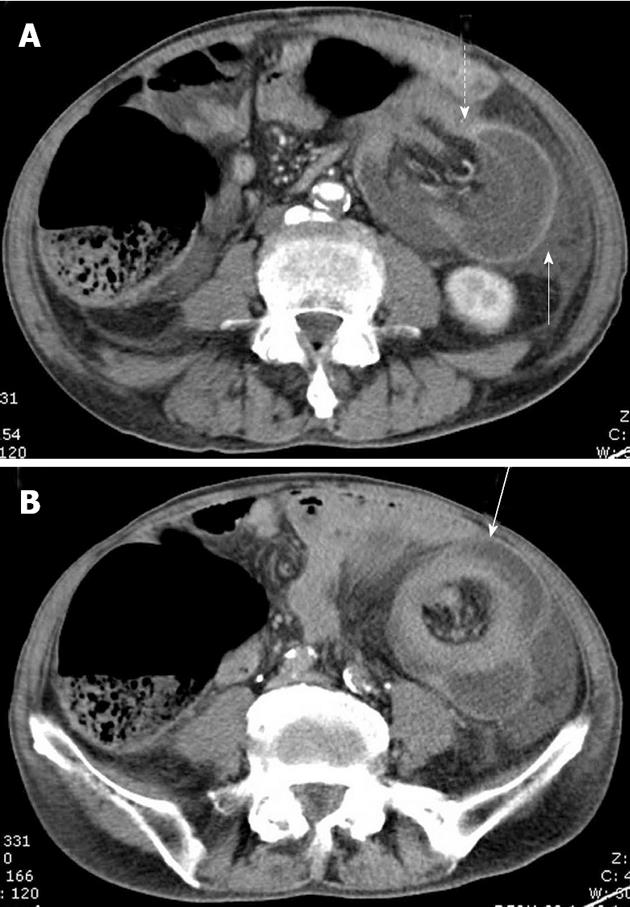
Figure 3 Abdominal computed tomography showing colocolic component of the intussusception (A, dashed arrow), thick-walled descending colon invaginated by transverse colon with mesenteric fat and vessels (A, solid arrow), and the typical “target sign” of colon within colon (B, solid arrow).
- Citation: Xu XQ, Hong T, Liu W, Zheng CJ, He XD, Li BL. A long adult intussusception secondary to transverse colon cancer. World J Gastroenterol 2013; 19(22): 3517-3519
- URL: https://www.wjgnet.com/1007-9327/full/v19/i22/3517.htm
- DOI: https://dx.doi.org/10.3748/wjg.v19.i22.3517


