
















©2013 Baishideng Publishing Group Co.
World J Gastroenterol. Jun 7, 2013; 19(21): 3358-3363
Published online Jun 7, 2013. doi: 10.3748/wjg.v19.i21.3358
Published online Jun 7, 2013. doi: 10.3748/wjg.v19.i21.3358
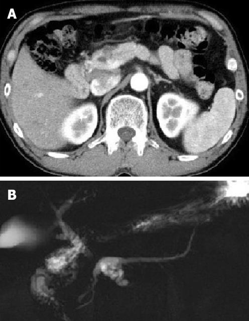
Figure 1 Computed tomography and magnetic resonance cholangiopancreatography imaging.
A: Multiple cystic lesions measuring 23 mm are indicated in the neck of the pancreas; B: Multiple cystic dilations of pancreatic branch duct connecting to the dilated main pancreatic duct are indicated in the neck of the pancreas.
- Citation: Hirabayashi K, Zamboni G, Ito H, Ogawa M, Kawaguchi Y, Yamashita T, Nakagohri T, Nakamura N. Synchronous pancreatic solid pseudopapillary neoplasm and intraductal papillary mucinous neoplasm. World J Gastroenterol 2013; 19(21): 3358-3363
- URL: https://www.wjgnet.com/1007-9327/full/v19/i21/3358.htm
- DOI: https://dx.doi.org/10.3748/wjg.v19.i21.3358