Copyright
©2013 Baishideng Publishing Group Co.
World J Gastroenterol. May 28, 2013; 19(20): 3157-3160
Published online May 28, 2013. doi: 10.3748/wjg.v19.i20.3157
Published online May 28, 2013. doi: 10.3748/wjg.v19.i20.3157
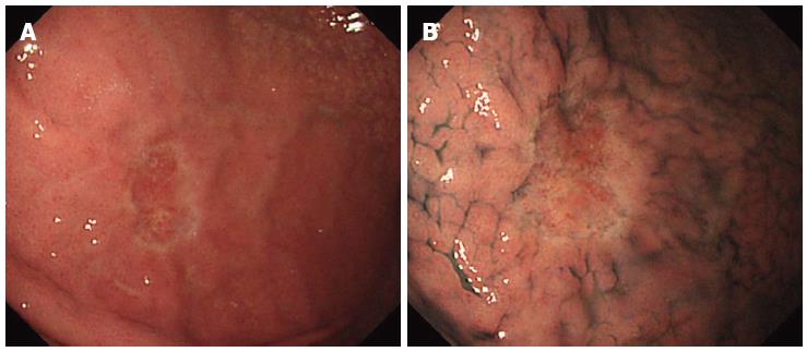
Figure 1 Pre-treatment endoscopic examination.
Esophagogastroduodenoscopy revealed pale depressed mucosal lesion on anterior wall of middle gastric body approximately 15 mm in size with no ulcerative finding and non-atrophic background mucosa. A: Conventional white light endoscopy; B: With indigo-carmine dye staining.
- Citation: Odagaki T, Suzuki H, Oda I, Yoshinaga S, Nonaka S, Katai H, Taniguchi H, Kushima R, Saito Y. Small undifferentiated intramucosal gastric cancer with lymph-node metastasis: Case report. World J Gastroenterol 2013; 19(20): 3157-3160
- URL: https://www.wjgnet.com/1007-9327/full/v19/i20/3157.htm
- DOI: https://dx.doi.org/10.3748/wjg.v19.i20.3157


