Copyright
©2013 Baishideng Publishing Group Co.
World J Gastroenterol. May 28, 2013; 19(20): 3108-3116
Published online May 28, 2013. doi: 10.3748/wjg.v19.i20.3108
Published online May 28, 2013. doi: 10.3748/wjg.v19.i20.3108
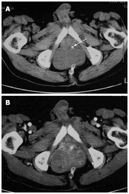
Figure 5 A 61-year-old man with a rectal gastrointestinal stromal tumor.
A: The mass located in the left wall of the rectum with fleck of calcification at the tumor margin (arrow); B: The mass enhanced heterogeneously following intravenous administration of contrast media.
- Citation: Jiang ZX, Zhang SJ, Peng WJ, Yu BH. Rectal gastrointestinal stromal tumors: Imaging features with clinical and pathological correlation. World J Gastroenterol 2013; 19(20): 3108-3116
- URL: https://www.wjgnet.com/1007-9327/full/v19/i20/3108.htm
- DOI: https://dx.doi.org/10.3748/wjg.v19.i20.3108


