Copyright
©2013 Baishideng Publishing Group Co.
World J Gastroenterol. May 28, 2013; 19(20): 3108-3116
Published online May 28, 2013. doi: 10.3748/wjg.v19.i20.3108
Published online May 28, 2013. doi: 10.3748/wjg.v19.i20.3108
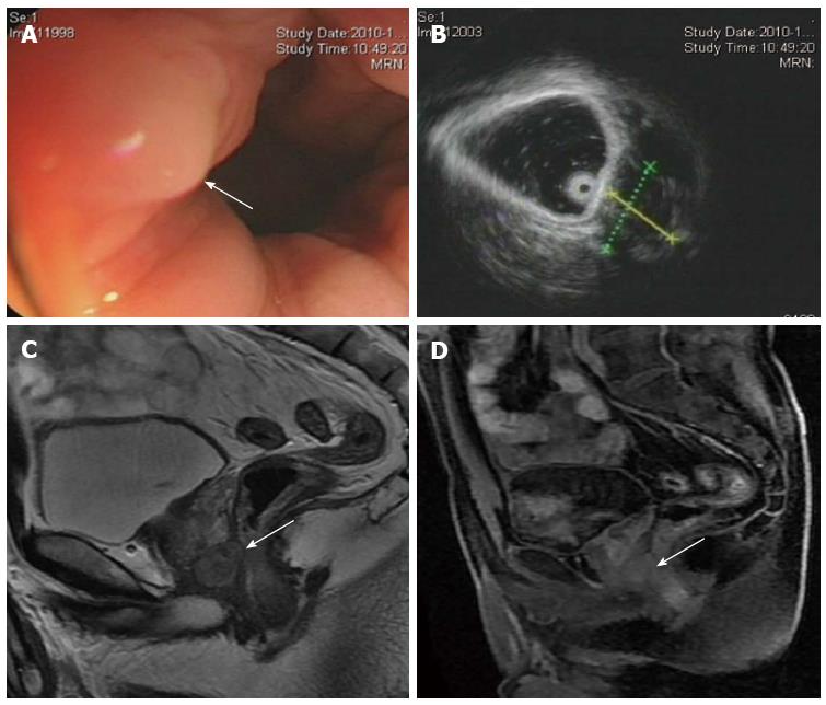
Figure 1 A 40-year-old man with a rectal gastrointestinal stromal tumor.
A: Colonoscopy shows a mass protruding from the rectal wall with intact overlying mucosa (arrow); B: Endoscopic ultrasonography shows a well-defined hypoechoic mass located along the right anterior aspect of the rectal wall; C: Sagittal T2-weighted magnetic resonance imaging shows an oval, homogenous, hyperintense mass with a sharp margin bordering the anterior rectal wall. A small area of anatomical continuity between the tumor and the anterior rectal wall is observed (arrow); D: Postcontrast T1-weighted image shows a slightly homogenously enhancing mass (arrow).
- Citation: Jiang ZX, Zhang SJ, Peng WJ, Yu BH. Rectal gastrointestinal stromal tumors: Imaging features with clinical and pathological correlation. World J Gastroenterol 2013; 19(20): 3108-3116
- URL: https://www.wjgnet.com/1007-9327/full/v19/i20/3108.htm
- DOI: https://dx.doi.org/10.3748/wjg.v19.i20.3108


