Copyright
©2013 Baishideng Publishing Group Co.
World J Gastroenterol. May 21, 2013; 19(19): 2935-2940
Published online May 21, 2013. doi: 10.3748/wjg.v19.i19.2935
Published online May 21, 2013. doi: 10.3748/wjg.v19.i19.2935
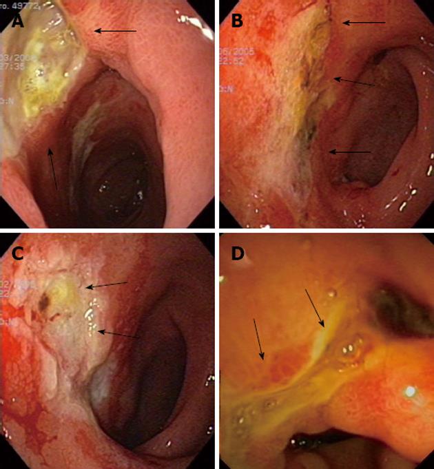
Figure 1 Figures of patient 5.
A: A big and deep ulcer was seen in the pyloric channel and duodenal bulb at diagnosis (arrows); B: The same ulcer as in (A) in the same location seen 3 mo later, partially healed (arrows); C: Ulcer located in the duodenal bulb (arrows) with an irregular and friable mucosa after 11 mo; D: Endoscopical view of antral, pyloric and duodenal bulb deformity (arrows) seen with endoscopic ultrasound scope 78 mo after diagnosis.
- Citation: Rodríguez-Lago I, Carretero C, Herráiz M, Subtil JC, Betés M, Rodríguez-Fraile M, Sola JJ, Bilbao JI, Muñoz-Navas M, Sangro B. Long-term follow-up study of gastroduodenal lesions after radioembolization of hepatic tumors. World J Gastroenterol 2013; 19(19): 2935-2940
- URL: https://www.wjgnet.com/1007-9327/full/v19/i19/2935.htm
- DOI: https://dx.doi.org/10.3748/wjg.v19.i19.2935


