Copyright
©2013 Baishideng Publishing Group Co.
World J Gastroenterol. May 21, 2013; 19(19): 2904-2912
Published online May 21, 2013. doi: 10.3748/wjg.v19.i19.2904
Published online May 21, 2013. doi: 10.3748/wjg.v19.i19.2904
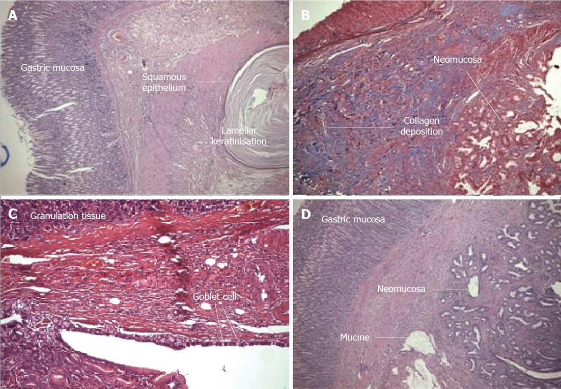
Figure 2 Histological and morphologic evaluation.
A: Unexpected neomucosal formation. The gastric corpus mucosa can be seen. The squamous epithelium and lamellar keratinization formed from the anastomosis [Hematoxylin and eosin (HE) × 100] (Group 3); B: Granulation tissue and newly formed neomucosa. The blue area is connective tissue (Masson Trichrome × 100) (Group 4); C: In the gastric mucosa of the large granulation tissue, newly formed goblet cells can be seen (HE × 100) (Group 4); D: The left side shows the gastric mucosa, and the right side shows newly formed neomucosa that contains mucin. The granulation tissue is reduced (HE × 100) (Group 4).
- Citation: Adas G, Adas M, Arikan S, Sarvan AK, Toklu AS, Mert S, Barut G, Kamali S, Koc B, Tutal F. Effect of growth hormone, hyperbaric oxygen and combined therapy on the gastric serosa. World J Gastroenterol 2013; 19(19): 2904-2912
- URL: https://www.wjgnet.com/1007-9327/full/v19/i19/2904.htm
- DOI: https://dx.doi.org/10.3748/wjg.v19.i19.2904


