
















©2013 Baishideng Publishing Group Co.
World J Gastroenterol. Apr 14, 2013; 19(14): 2187-2196
Published online Apr 14, 2013. doi: 10.3748/wjg.v19.i14.2187
Published online Apr 14, 2013. doi: 10.3748/wjg.v19.i14.2187
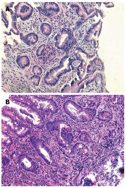
Figure 2 Representative hematoxylin and eosin staining of gastric tissue from chronic atrophic gastritis (A) and duodenogastric reflux (B).
Isolated metaplasia of glandular epithelium and mild inflammation of the lamina propria was found in the tissue of duodenogastric reflux patients (original magnification, × 200).
- Citation: Chen TF, Yadav PK, Wu RJ, Yu WH, Liu CQ, Lin H, Liu ZJ. Comparative evaluation of intragastric bile acids and hepatobiliary scintigraphy in the diagnosis of duodenogastric reflux. World J Gastroenterol 2013; 19(14): 2187-2196
- URL: https://www.wjgnet.com/1007-9327/full/v19/i14/2187.htm
- DOI: https://dx.doi.org/10.3748/wjg.v19.i14.2187