Copyright
©2013 Baishideng Publishing Group Co.
World J Gastroenterol. Apr 7, 2013; 19(13): 2037-2043
Published online Apr 7, 2013. doi: 10.3748/wjg.v19.i13.2037
Published online Apr 7, 2013. doi: 10.3748/wjg.v19.i13.2037
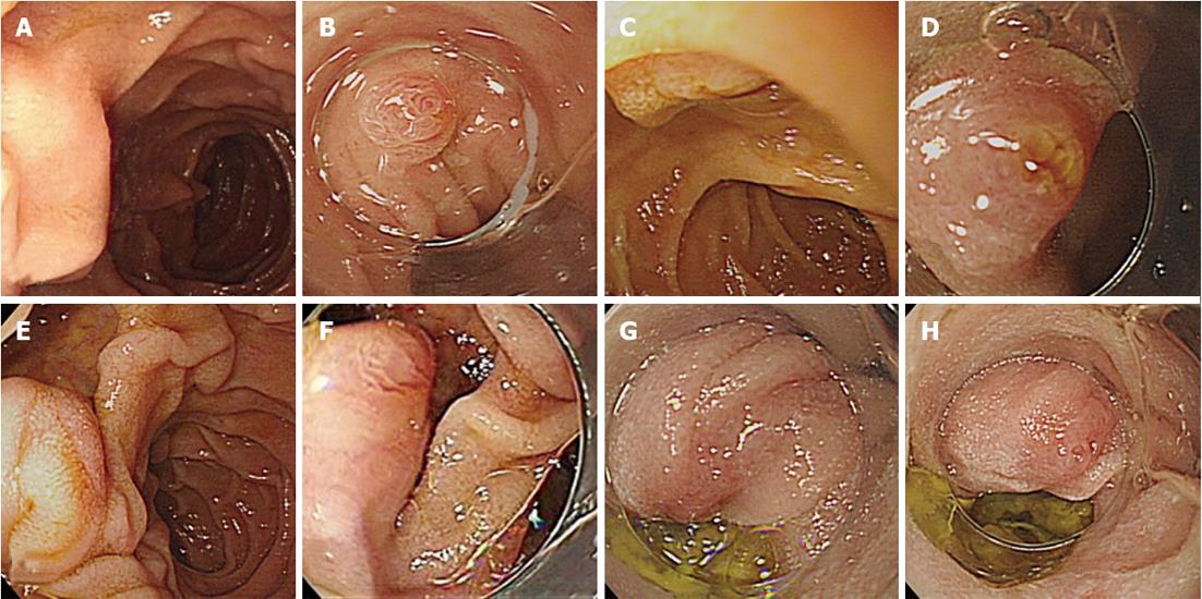
Figure 4 Incomplete observation of the ampulla of Vater by conventional endoscopy and outcomes of cap-assisted endoscopy.
A: Incomplete observation of the ampulla of Vater (AV) by forward-viewing endoscopy due to a folded mucous membrane; C: Incomplete observation of the AV due to the close proximity of the endoscope tip to a superior ampulla lesion; E: Incomplete observations of the AV on the edge of diverticulum; B, D, F: Complete observation of the AV, including the orifice, by short cap-assisted endoscopy (A→B, C→D, E→F); G: Incomplete observation of the AV by short cap-assisted endoscopy due to a loop in the scope; H: Complete observation of the AV by long cap-assisted endoscopy (G→H).
- Citation: Choi YR, Han JH, Cho YS, Han HS, Chae HB, Park SM, Youn SJ. Efficacy of cap-assisted endoscopy for routine examining the ampulla of Vater. World J Gastroenterol 2013; 19(13): 2037-2043
- URL: https://www.wjgnet.com/1007-9327/full/v19/i13/2037.htm
- DOI: https://dx.doi.org/10.3748/wjg.v19.i13.2037


