Copyright
©2013 Baishideng Publishing Group Co.
World J Gastroenterol. Mar 14, 2013; 19(10): 1652-1656
Published online Mar 14, 2013. doi: 10.3748/wjg.v19.i10.1652
Published online Mar 14, 2013. doi: 10.3748/wjg.v19.i10.1652
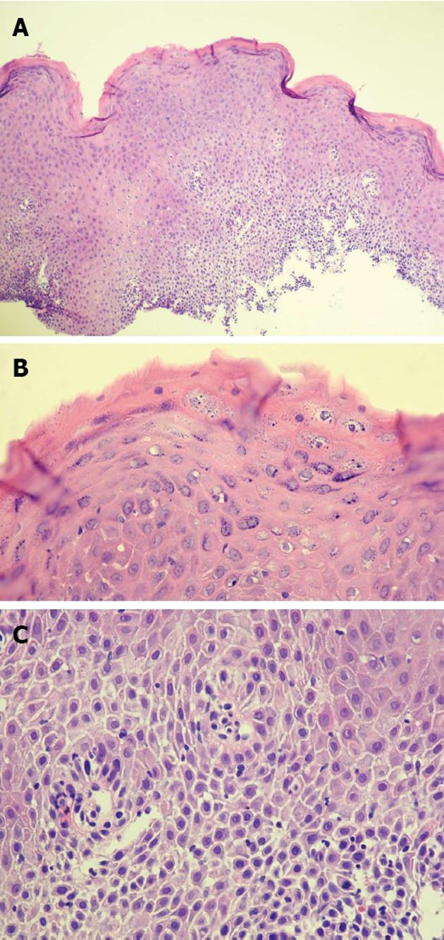
Figure 3 Chronic inflammatory cell infiltration of the epithelium was also present and was mainly composed of lymphocytes.
A: Low power view of upper esophagus shows extensive severe keratinization, organizational disarray of cell arrangement and spongiosis of lower layer associated with inflammatory infiltration. (E, × 40); B: High power of parakeratotic cells and accumulation of keratohyaline granules in the cytoplasm of the mature keratinocytes. In this zone keratinization is mild. (HE, × 400); C: High power view of spongiosis of lower layers of squamous epithelium associated with sprinkling of lymphocytes typical of longstanding chronic inflammation. (HE, × 400).
- Citation: Ynson ML, Forouhar F, Vaziri H. Case report and review of esophageal lichen planus treated with fluticasone. World J Gastroenterol 2013; 19(10): 1652-1656
- URL: https://www.wjgnet.com/1007-9327/full/v19/i10/1652.htm
- DOI: https://dx.doi.org/10.3748/wjg.v19.i10.1652


