Copyright
©2013 Baishideng Publishing Group Co.
World J Gastroenterol. Jan 7, 2013; 19(1): 129-132
Published online Jan 7, 2013. doi: 10.3748/wjg.v19.i1.129
Published online Jan 7, 2013. doi: 10.3748/wjg.v19.i1.129
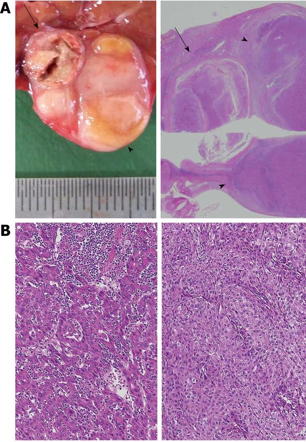
Figure 2 Macroscopic findings of the resected specimen, calcified and non-calcified lesions.
A: Left, macroscopic examination showed a 10-mm tumor with ring calcification (arrow) and an 18-mm tumor (arrowhead) existing side-by-side and encapsulated within the same thick fibrous tissue; Right, a prepared specimen (loupe image, hematoxylin and eosin staining); B: Left, microscopic examination findings of the calcified lesion showed the poorly differentiated hepatocellular carcinoma (hematoxylin and eosin staining; original magnification, × 200); Right, microscopic examination findings of the non-calcified lesion showed moderately differentiated hepatocellular carcinoma (hematoxylin and eosin staining; original magnification, × 200).
- Citation: Murakami T, Morioka D, Takakura H, Miura Y, Togo S. Small hepatocellular carcinoma with ring calcification: A case report and literature review. World J Gastroenterol 2013; 19(1): 129-132
- URL: https://www.wjgnet.com/1007-9327/full/v19/i1/129.htm
- DOI: https://dx.doi.org/10.3748/wjg.v19.i1.129


