Copyright
©2012 Baishideng Publishing Group Co.
World J Gastroenterol. Feb 7, 2012; 18(5): 435-444
Published online Feb 7, 2012. doi: 10.3748/wjg.v18.i5.435
Published online Feb 7, 2012. doi: 10.3748/wjg.v18.i5.435
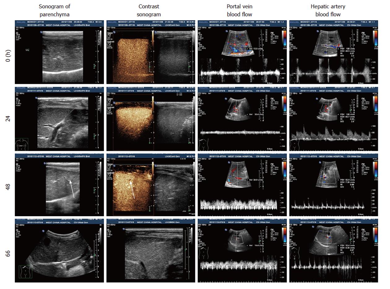
Figure 3 Liver sonogram of the Macaca mulatta model at different times.
Arrows show a slightly enhanced echo and lower perfusion of SonoVue at 48 h.
-
Citation: Zhou P, Xia J, Guo G, Huang ZX, Lu Q, Li L, Li HX, Shi YJ, Bu H. A
Macaca mulatta model of fulminant hepatic failure. World J Gastroenterol 2012; 18(5): 435-444 - URL: https://www.wjgnet.com/1007-9327/full/v18/i5/435.htm
- DOI: https://dx.doi.org/10.3748/wjg.v18.i5.435


