Copyright
©2012 Baishideng Publishing Group Co.
World J Gastroenterol. Dec 14, 2012; 18(46): 6720-6728
Published online Dec 14, 2012. doi: 10.3748/wjg.v18.i46.6720
Published online Dec 14, 2012. doi: 10.3748/wjg.v18.i46.6720
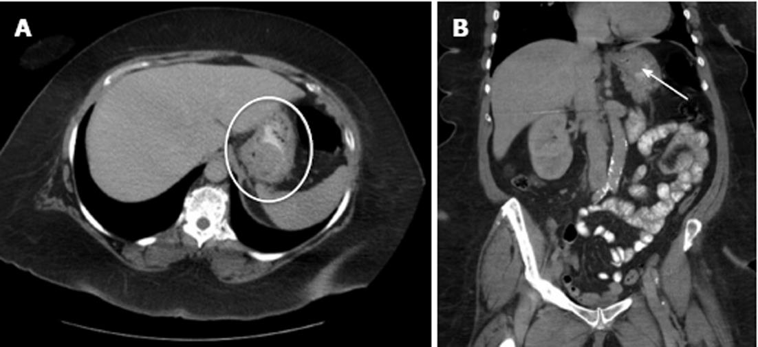
Figure 3 Gastroesophageal junction gastrointestinal stromal tumors.
A: An axial computed tomography image of a gastric gastrointestinal stromal tumor (white oval) located along the posterior wall of the gastroesophageal junction (GEJ); B: Coronal images of the tumor (white arrow) show its proximity to the GEJ.
- Citation: Roggin KK, Posner MC. Modern treatment of gastric gastrointestinal stromal tumors. World J Gastroenterol 2012; 18(46): 6720-6728
- URL: https://www.wjgnet.com/1007-9327/full/v18/i46/6720.htm
- DOI: https://dx.doi.org/10.3748/wjg.v18.i46.6720


