Copyright
©2012 Baishideng Publishing Group Co.
World J Gastroenterol. Dec 7, 2012; 18(45): 6651-6656
Published online Dec 7, 2012. doi: 10.3748/wjg.v18.i45.6651
Published online Dec 7, 2012. doi: 10.3748/wjg.v18.i45.6651
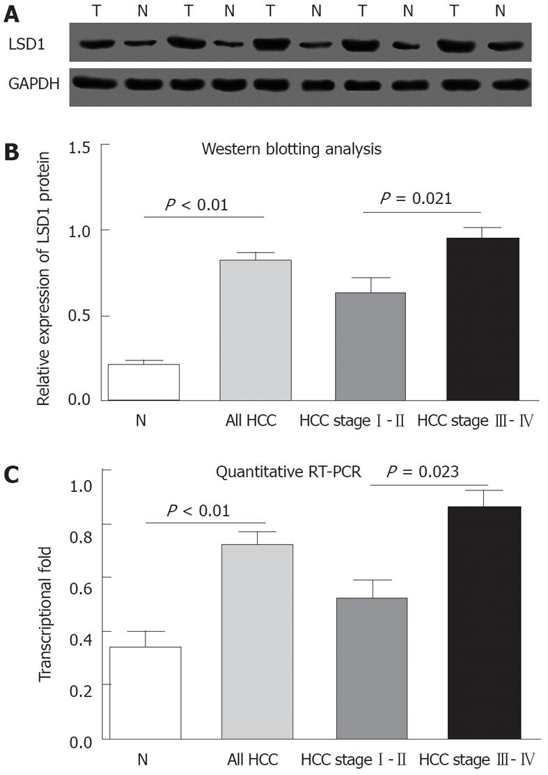
Figure 2 Increased lysine specific demethylase 1 protein and mRNA levels in hepatocellular carcinoma with different tumor node metastasis stages and adjacent non-neoplastic liver tissues.
A: Representative Western blotting of lysine specific demethylase 1 (LSD1) protein levels in hepatocellular carcinoma (HCC) tissues (T) and adjacent non-neoplastic liver tissues (N); B: Semiquantitative Western blotting showed that the expression levels of LSD1 protein were significantly higher than those in adjacent non-neoplastic liver tissues (P < 0.01). Additionally, the expression levels of LSD1 protein increased with ascending tumor node metastasis (TNM) stages. Glyceraldehyde-3-phosphate dehydrogenase (GAPDH) was used as an internal control. P values, mean and SD were given (t test); C: Quantitative reverse transcription-polymerase chain reaction (RT-PCR) assay showed significantly increased LSD1 mRNA levels in HCC tissues compared with adjacent non-neoplastic liver tissues (P < 0.01). Additionally, the expression levels of LSD1 mRNA were increased with ascending tumor TNM stages. GAPDH was used as the internal control. P values, mean and SD were given (Mann-Whitney test).
- Citation: Zhao ZK, Yu HF, Wang DR, Dong P, Chen L, Wu WG, Ding WJ, Liu YB. Overexpression of lysine specific demethylase 1 predicts worse prognosis in primary hepatocellular carcinoma patients. World J Gastroenterol 2012; 18(45): 6651-6656
- URL: https://www.wjgnet.com/1007-9327/full/v18/i45/6651.htm
- DOI: https://dx.doi.org/10.3748/wjg.v18.i45.6651


