Copyright
©2012 Baishideng Publishing Group Co.
World J Gastroenterol. Nov 7, 2012; 18(41): 5990-5993
Published online Nov 7, 2012. doi: 10.3748/wjg.v18.i41.5990
Published online Nov 7, 2012. doi: 10.3748/wjg.v18.i41.5990
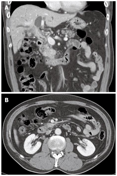
Figure 4 Follow up abdominal computed tomography scan.
A: Coronal image revealed a stenosis of the common hepatic and the proximal common bile duct (white arrow) with significant thickening and inner wall enhancement of the bile duct; B: There was no pancreatic relapse (white arrow).
- Citation: Kim JH, Chang JH, Nam SM, Lee MJ, Maeng IH, Park JY, Im YS, Kim TH, Kim CW, Han SW. Newly developed autoimmune cholangitis without relapse of autoimmune pancreatitis after discontinuing prednisolone. World J Gastroenterol 2012; 18(41): 5990-5993
- URL: https://www.wjgnet.com/1007-9327/full/v18/i41/5990.htm
- DOI: https://dx.doi.org/10.3748/wjg.v18.i41.5990


