Copyright
©2012 Baishideng Publishing Group Co.
World J Gastroenterol. Oct 21, 2012; 18(39): 5640-5644
Published online Oct 21, 2012. doi: 10.3748/wjg.v18.i39.5640
Published online Oct 21, 2012. doi: 10.3748/wjg.v18.i39.5640
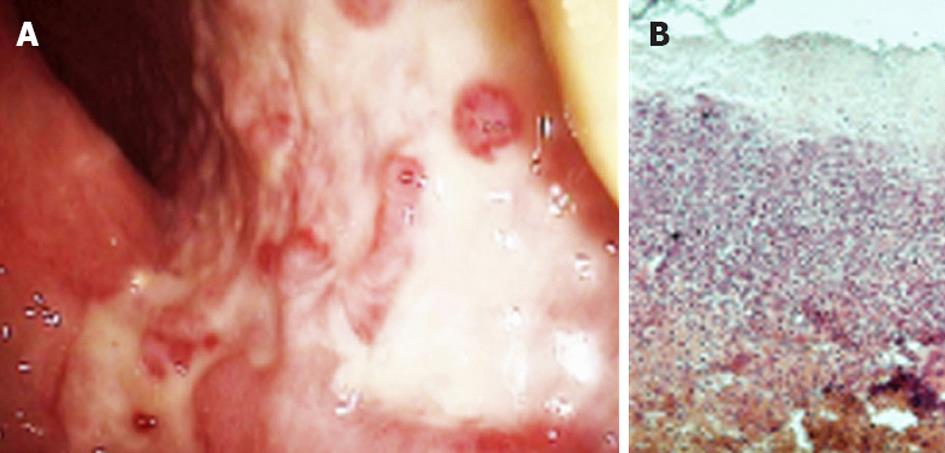
Figure 3 Morphologic changes confirming ischemic colitis by lower digestive endoscopy and histology.
A: Areas of mucosal ulceration, pseudomembranes and subsequent granulation observed at colonoscopy; B: Biopsy specimens from affected areas showing submucosal necrosis, inflammatory infiltration, cryptal atrophy and hyalinization. Hematoxylin-eosin staining, ×100. Courtesy Dr. Simionescu.
- Citation: Georgescu EF, Carstea D, Dumitrescu D, Teodorescu R, Carstea A. Ischemic colitis and large bowel infarction: A case report. World J Gastroenterol 2012; 18(39): 5640-5644
- URL: https://www.wjgnet.com/1007-9327/full/v18/i39/5640.htm
- DOI: https://dx.doi.org/10.3748/wjg.v18.i39.5640


