Copyright
©2012 Baishideng Publishing Group Co.
World J Gastroenterol. Oct 21, 2012; 18(39): 5640-5644
Published online Oct 21, 2012. doi: 10.3748/wjg.v18.i39.5640
Published online Oct 21, 2012. doi: 10.3748/wjg.v18.i39.5640
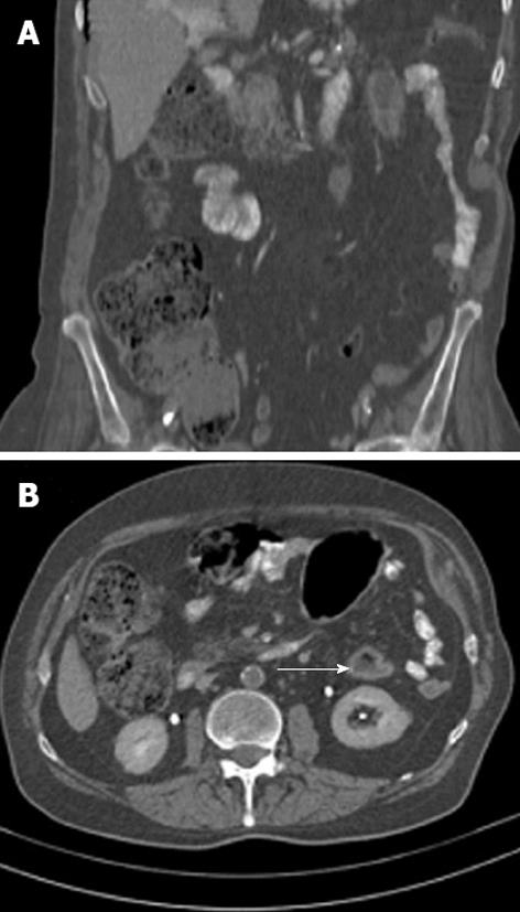
Figure 2 Contrast-enhanced abdominal computed tomography scans suggesting segmental colitis involving the splenic flexure and the descending colon.
A: Coronal sections with zones of mural thickening of the colon just below the splenic flexure and a long and narrow axial stenosis of the descending colon; B: Sagittal sections showing (arrow) the "double halo" sign, a tomographic equivalent of the classic “thumbprinting” that is observed in barium enemas.
- Citation: Georgescu EF, Carstea D, Dumitrescu D, Teodorescu R, Carstea A. Ischemic colitis and large bowel infarction: A case report. World J Gastroenterol 2012; 18(39): 5640-5644
- URL: https://www.wjgnet.com/1007-9327/full/v18/i39/5640.htm
- DOI: https://dx.doi.org/10.3748/wjg.v18.i39.5640


