Copyright
©2012 Baishideng Publishing Group Co.
World J Gastroenterol. Sep 14, 2012; 18(34): 4704-4713
Published online Sep 14, 2012. doi: 10.3748/wjg.v18.i34.4704
Published online Sep 14, 2012. doi: 10.3748/wjg.v18.i34.4704
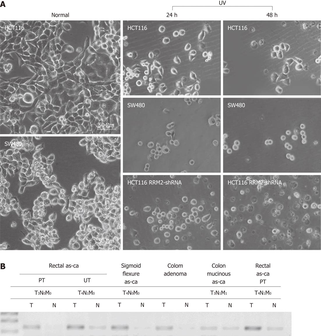
Figure 5 Light-microscopic photograph of cellular morphology.
A: After 48 h of culture in normal conditions or exposed to ultraviolet (UV), HCT116 cells [high expression level of ribonucleotide reductase M2 (RRM2)] and SW480 cells (low expression level of RRM2) were photographed (20 × magnification). With UV-C irradiating, SW480 cells formed spheroids and HCT116 cells still adhered to each other and attached; B: Expression of RRM2 mRNA in clinical tissue specimens. Polymerase chain reaction analyses of 25 colorectal cancer patients (we show 6 patients) showed the expression levels of RRM2. PT: Protrude type; UT: Ulcerative type; as-ca: Adenocarcinoma.
- Citation: Lu AG, Feng H, Wang PXZ, Han DP, Chen XH, Zheng MH. Emerging roles of the ribonucleotide reductase M2 in colorectal cancer and ultraviolet-induced DNA damage repair. World J Gastroenterol 2012; 18(34): 4704-4713
- URL: https://www.wjgnet.com/1007-9327/full/v18/i34/4704.htm
- DOI: https://dx.doi.org/10.3748/wjg.v18.i34.4704


