Copyright
©2012 Baishideng Publishing Group Co.
World J Gastroenterol. Aug 21, 2012; 18(31): 4228-4232
Published online Aug 21, 2012. doi: 10.3748/wjg.v18.i31.4228
Published online Aug 21, 2012. doi: 10.3748/wjg.v18.i31.4228
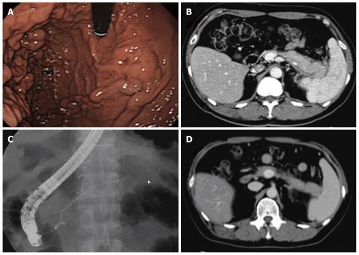
Figure 2 The splenic vein was not reperfused, although the autoimmune pancreatitis improved.
A: Esophagogastoroduodenoscopy showed gastric varices in the fundus of the stomach; B: Computed tomography showed a locally enlarged pancreatic tail with a capsule-like rim, an obstructed splenic vein, and splenomegaly; C: Endoscopic retrograde cholangiopancreatography showed irregular narrowing of the main pancreatic duct in the pancreatic tail (arrowhead); D: Autoimmune pancreatitis improved following steroid therapy, but the splenic vein was not reperfused.
- Citation: Goto N, Mimura J, Itani T, Hayashi M, Shimada Y, Matsumori T. Autoimmune pancreatitis complicated by gastric varices: A report of 3 cases. World J Gastroenterol 2012; 18(31): 4228-4232
- URL: https://www.wjgnet.com/1007-9327/full/v18/i31/4228.htm
- DOI: https://dx.doi.org/10.3748/wjg.v18.i31.4228


