Copyright
©2012 Baishideng Publishing Group Co.
World J Gastroenterol. Aug 21, 2012; 18(31): 4127-4135
Published online Aug 21, 2012. doi: 10.3748/wjg.v18.i31.4127
Published online Aug 21, 2012. doi: 10.3748/wjg.v18.i31.4127
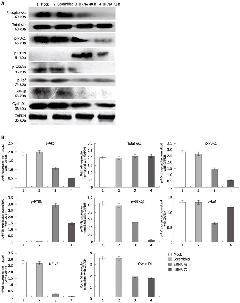
Figure 3 Expression of proteins in esophageal squamous cancer cells TE13 and compared with control.
A: Expression analysis of protein kinase B (pAkt), total Akt, protein Glycogen synthase kinase 3β (pGSK3β), pRaf, protein Phosphoinositide-dependent kinase-1 (pPDK1), phosphatase and tensin homolog (pPTEN), nuclear factor-κB (NF-κB) and cyclin D1 proteins compared in esophageal squamous cancer cells TE13. Lane 1: Mock; Lane 2: Scrambled (non-target siRNA); Lane 3: TC21 siRNA transfected for 48 h; Lane 4: TC21 siRNA transfected for 72 h; B: Bar diagram showing relative levels of proteins in comparison with control protein GAPDH. GAPDH: Glyceraldehyde-3-phosphate dehydrogenase.
- Citation: Hasan MR, Chauhan SS, Sharma R, Ralhan R. siRNA-mediated downregulation of TC21 sensitizes esophageal cancer cells to cisplatin. World J Gastroenterol 2012; 18(31): 4127-4135
- URL: https://www.wjgnet.com/1007-9327/full/v18/i31/4127.htm
- DOI: https://dx.doi.org/10.3748/wjg.v18.i31.4127


