Copyright
©2012 Baishideng Publishing Group Co.
World J Gastroenterol. Aug 21, 2012; 18(31): 4102-4117
Published online Aug 21, 2012. doi: 10.3748/wjg.v18.i31.4102
Published online Aug 21, 2012. doi: 10.3748/wjg.v18.i31.4102
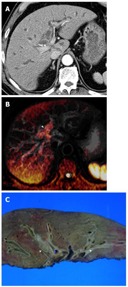
Figure 13 Hilar cholangiocarcinoma (intraductal-growing type) in a 67-year-old man.
A: Contrast-enhanced CT shows mild intrahepatic duct dilatation. However, intraductal masses are not clearly depicted on CT; B: Fusion image of T2-weighted and diffusion-weighted images at b = 800 s/mm2 shows high signal intensity (arrowhead) at the first branch of the intrahepatic duct; C: Photograph of the gross specimen shows intraductal growing masses (arrowheads) in the bile duct. Histological analysis revealed biliary intraepithelial neoplasia with high grade dysplasia. CT: Computed tomography.
- Citation: Lee NK, Kim S, Kim GH, Kim DU, Seo HI, Kim TU, Kang DH, Jang HJ. Diffusion-weighted imaging of biliopancreatic disorders: Correlation with conventional magnetic resonance imaging. World J Gastroenterol 2012; 18(31): 4102-4117
- URL: https://www.wjgnet.com/1007-9327/full/v18/i31/4102.htm
- DOI: https://dx.doi.org/10.3748/wjg.v18.i31.4102


