Copyright
©2012 Baishideng Publishing Group Co.
World J Gastroenterol. Aug 14, 2012; 18(30): 4051-4058
Published online Aug 14, 2012. doi: 10.3748/wjg.v18.i30.4051
Published online Aug 14, 2012. doi: 10.3748/wjg.v18.i30.4051
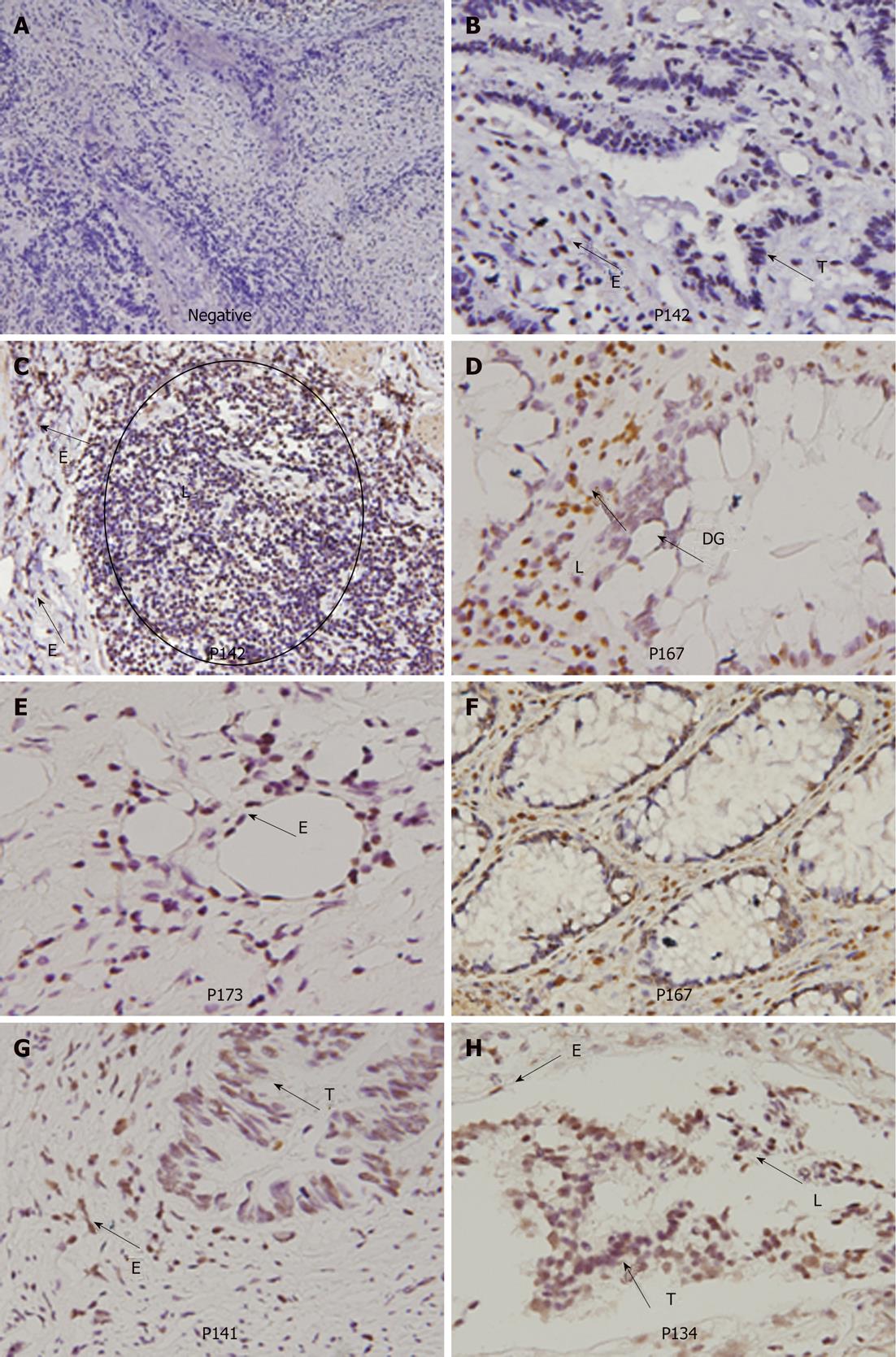
Figure 3 Immunohistochemical analysis of human papilloma virus 16 E6 protein in colorectal tumors and adjacent normal tissues.
A: A negative result of immunostaining in tumor cells (100 ×); B: Human papilloma virus 16 (HPV16) E6 protein expressed in endothelial cells (400 ×); C: HPV16 E6 protein expressed in endothelial cells and lymphocytes (100 ×); D: HPV16 E6 protein expressed in lymphocytes and dysplastic gland (400 ×); E: HPV16 E6 protein expressed in endothelial cells and tumor cells (400 ×); F: HPV16 E6 protein expressed in normal gland (400 ×); G: HPV16 E6 protein in endothelial cells and Fibroblast (400 ×); H: HPV16 E6 protein in endothelial cells, lymphocytes and tumor cells (400 ×). E: Endothelial cells; T: Tumor cells; L: Lymphocytes; DG: Dysplastic gland.
- Citation: Chen TH, Huang CC, Yeh KT, Chang SH, Chang SW, Sung WW, Cheng YW, Lee H. Human papilloma virus 16 E6 oncoprotein associated with p53 inactivation in colorectal cancer. World J Gastroenterol 2012; 18(30): 4051-4058
- URL: https://www.wjgnet.com/1007-9327/full/v18/i30/4051.htm
- DOI: https://dx.doi.org/10.3748/wjg.v18.i30.4051


