Copyright
©2012 Baishideng Publishing Group Co.
World J Gastroenterol. Aug 14, 2012; 18(30): 3977-3991
Published online Aug 14, 2012. doi: 10.3748/wjg.v18.i30.3977
Published online Aug 14, 2012. doi: 10.3748/wjg.v18.i30.3977
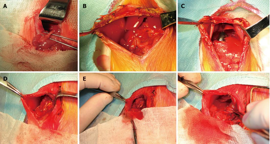
Figure 1 Images of experimental obstructive jaundice and internal biliary drainage.
A: Dissection revealing the common bile duct; B: Reoperation after 3 d of common bile duct ligation. Light yellow abdominal ascites were present in the right side of the abdominal cavity; C: The proximal bile duct showed a remarkable expansion (dark blue color) after 3 d of common bile duct ligation; D: The PE-10 polyethylene tube was positioned with the end tied in the right hepatorenal recess; E: Brown bile flowed out while the catheter end was open; F: The distal 3 cm segment of the catheter was inserted into the duodenum for internal biliary drainage.
-
Citation: Zhou YK, Qin HL, Zhang M, Shen TY, Chen HQ, Ma YL, Chu ZX, Zhang P, Liu ZH. Effects of
Lactobacillus plantarum on gut barrier function in experimental obstructive jaundice. World J Gastroenterol 2012; 18(30): 3977-3991 - URL: https://www.wjgnet.com/1007-9327/full/v18/i30/3977.htm
- DOI: https://dx.doi.org/10.3748/wjg.v18.i30.3977


