Copyright
©2012 Baishideng Publishing Group Co.
World J Gastroenterol. Aug 7, 2012; 18(29): 3875-3882
Published online Aug 7, 2012. doi: 10.3748/wjg.v18.i29.3875
Published online Aug 7, 2012. doi: 10.3748/wjg.v18.i29.3875
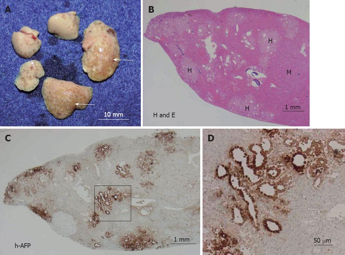
Figure 1 Macro- and microscopic images of the liver from group A mice.
A: The urokinase-type plasminogen activator/severe combined immunodeficient mouse mice were transplanted with human gastric cancer cells (h-GCCs) and euthanized 56 d later, at which time the livers were isolated and photographed; B: The arrows in A point to concentrated regions of h-GCC colonies, and the sections were stained with hematoxylin and eosin (H and E). H and M in B represent h-GCC colonies and m-liver cell regions, respectively; C: The sections were stained with anti-human alpha-fetoprotein (h-AFP) antibodies; D: The square region in C is enlarged and shown.
-
Citation: Fujiwara S, Fujioka H, Tateno C, Taniguchi K, Ito M, Ohishi H, Utoh R, Ishibashi H, Kanematsu T, Yoshizato K. A novel animal model for
in vivo study of liver cancer metastasis. World J Gastroenterol 2012; 18(29): 3875-3882 - URL: https://www.wjgnet.com/1007-9327/full/v18/i29/3875.htm
- DOI: https://dx.doi.org/10.3748/wjg.v18.i29.3875


