Copyright
©2012 Baishideng Publishing Group Co.
World J Gastroenterol. Jul 14, 2012; 18(26): 3451-3457
Published online Jul 14, 2012. doi: 10.3748/wjg.v18.i26.3451
Published online Jul 14, 2012. doi: 10.3748/wjg.v18.i26.3451
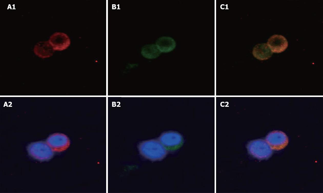
Figure 1 Localization of uncoupling protein-2 and glucagon-like peptide-1 in NCI-H716 cells.
Immunofluorescence staining in human NCI-H716 cells which serve as a model for L-cells. A1, A2: Glucagon-like peptide-1 (GLP-1) antibody staining (red); B1, B2: Uncoupling protein-2 (UCP2) antibody staining (green); C1, C2: Merged image shows co-expression of UCP2 and GLP-1. Blue (Hoechst) staining indicates the nuclei. Original magnification, ¡Á 400.
- Citation: Chen Y, Li ZY, Yang Y, Zhang HJ. Uncoupling protein 2 regulates glucagon-like peptide-1 secretion in L-cells. World J Gastroenterol 2012; 18(26): 3451-3457
- URL: https://www.wjgnet.com/1007-9327/full/v18/i26/3451.htm
- DOI: https://dx.doi.org/10.3748/wjg.v18.i26.3451


